

Melanoma in Situ
Melanoma in situ does not have a single discoverer because it is considered the earliest stage of melanoma rather than a separate disease. Melanoma itself was first described in 1804 by French physician René Théophile Hyacinthe Laennec, and later, in 1820, William Norris documented its ability to spread and suggested that it could run in families. The term "melanoma in situ" became widely used during the twentieth century as pathologists recognized that melanoma could be confined to the epidermis before invading deeper layers of the skin.
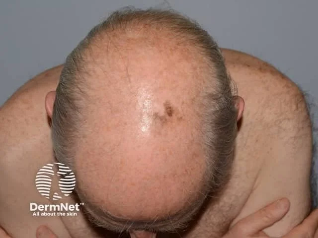
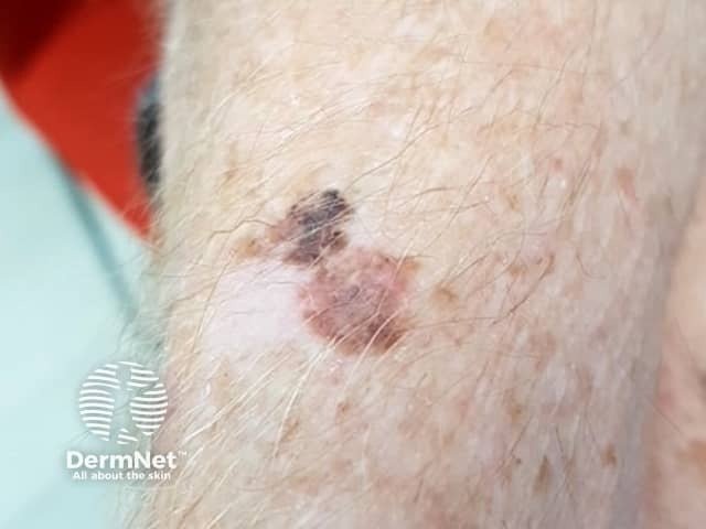
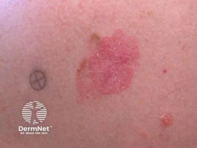
Melanoma in situ can develop anywhere on the skin but is most commonly found on sun-exposed areas, including the face, neck, back, chest, arms, and legs. In men, it most often occurs on the back and trunk, while in women it is more commonly found on the legs. A subtype called Lentigo maligna usually develops on the face and other areas that have experienced many years of sun exposure.
There is no standard size for melanoma in situ because it varies depending on when it is detected. Most lesions measure between 5 and 15 millimeters (0.5–1.5 centimeters or about 0.2–0.6 inches) in diameter at diagnosis, although some may be smaller or larger. Because the cancer is confined to the epidermis, it has not yet invaded the deeper layers of the skin.
The primary cause of melanoma in situ is damage to melanocytes caused by ultraviolet (UV) radiation from sunlight or indoor tanning beds. UV radiation causes genetic mutations in pigment-producing cells, allowing abnormal cells to develop within the epidermis before they invade deeper tissues.
Other factors that increase the risk of melanoma in situ include fair skin, light-colored eyes, blond or red hair, a history of severe sunburns, numerous or atypical moles, a family history of melanoma, inherited genetic mutations such as CDKN2A mutations, older age, and a weakened immune system. Although these factors increase the risk, melanoma in situ can develop in people of any skin color.
Melanoma in situ is most commonly diagnosed in adults over the age of 50, although it can occur in younger adults, especially those with significant ultraviolet exposure or a strong family history of melanoma. The risk increases with age due to cumulative sun damage.
The standard treatment for melanoma in situ is surgical excision with appropriate margins to ensure complete removal of the abnormal cells. Mohs micrographic surgery or staged excision may be recommended for lesions on the face or other cosmetically sensitive areas. In selected patients who are not good candidates for surgery, treatments such as topical Imiquimod or radiation therapy may be considered, although surgery remains the preferred treatment.
Melanoma in situ is generally not deadly because the abnormal melanocytes are confined to the epidermis and have not spread into deeper tissues or other parts of the body. When treated promptly with complete surgical removal, the cure rate exceeds 99%. However, if left untreated, melanoma in situ can progress to invasive melanoma, which can become life-threatening.
Visual Images
〰️
Visual Images 〰️


Medical references (source)
American Academy of Dermatology. (2024). Melanoma: Overview.
American Cancer Society. (2024). Melanoma skin cancer.
Mackie, R. M. (2003). History of melanoma. The Lancet, 361(9367), 571.
National Cancer Institute. (2024). Melanoma treatment (PDQ®).
National Comprehensive Cancer Network. (2025). NCCN Clinical Practice Guidelines in Oncology: Cutaneous Melanoma.